Georgetown University Medical Center

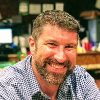
Mark Burns, PhD
Professor Ph.D., 2000, National University of Ireland, Galway, IrelandNew Research Building, Room EG17A Phone: 202.687.4735 Email: [email protected]
Dr Burns’ lab investigates the link between traumatic brain injury (TBI) and Alzheimer’s disease. Exposure to TBI can quadruple the risk of developing Alzheimer’s disease, and amyloid plaques similar to those seen in Alzheimer’s disease have been found in the brain of TBI fatalities. Dr Burns’ lab recently found that the same pathways that are activated long-term in Alzheimer’s disease are activated short-term after TBI. By blocking the activation of these pathways in mice, the physical disability or memory impairments following brain trauma were completely abolished and the amount of brain damage was reduced by over 70%. This research is providing new insights into how toxic proteins produced in the brain in Alzheimer’s disease and TBI are causing neuronal cell damage and memory loss.
First of its Kind Study Explains Why Rest is Critical After a Concussion
“‘The findings mirror what has been observed about such damage in humans years after a brain injury, especially among athletes,’ Burns says. ‘Studies have shown that almost all people with single concussions spontaneously recover, but athletes who play contact sports are much more susceptible to lasting brain damage. These findings help fill in the picture of how and when concussions and mild head trauma can lead to sustained brain damage.’”
Female and Male Mice Suffer, Recover from TBI Differently
“The researchers focused on how sex alters key neuroinflammatory responses that follow TBI. They specifically looked at microglial cells, which are the resident immune cells of the brain, and movement of macrophages from the blood into the injured brain. Macrophages, which are also immune cells, offer the first line of defense against infection.”
“Burns also says that although the female mice have less of the negative effects of neuroinflammation such as neuron cell death, there are also positive aspects to neuroinflammation that are missing in female mice such as waste removal and wound healing. Understanding how to minimize the negative effects while maximizing the positive effects of inflammation is an important goal in TBI research.”

We've published..
Last updated June 2024
Mokbel, AY, Burns MP, Main BS (2024) The contribution of the meningeal immune interface to neuroinflammation in traumatic brain injury. Journal of Neuroinflammation 21: 135 doi.org/10.1186/s12974-024-03122-7
Chapman DP, Powers SD, ^ Vicini S, ^ Ryan TJ, ^ Burns MP (^joint senior authors) (2024) Amnesia after repeated head impact is caused by impaired synaptic plasticity in the memory engram. Journal of Neuroscience 44 (8) e1560232024 doi.org/10.1523/JNEUROSCI.1560-23.2024
Buenaventura RG, Harvey AC, Burns MP, Main BS (202 3 ) Traumatic brain injury induces an adaptive immune response in the meningeal transcriptome that is amplified by aging . Frontiers in Neuroscience 17: 1210175
doi: 10.3389/fnins.2023.1210175
Romariz SAA, Main BS, Harvey AC, Longo BM, Burns MP (2023) Delayed treatment with ceftriaxone reverses the enhanced sensitivity of TBI mice to chemically-induced seizures. PLoS O ne 18(7): e0288363. https://doi.org/10.1371/journal.pone.0288363
Chapman DP, Vicni S, Burns MP, Evans R (2023) Single Neuron Modeling Identifies Potassium Channel Modulation as Potential Target for Repetitive head Impacts. Neuroinformatics https://pubmed.ncbi.nlm.nih.gov/37294503/ . (Click here for data and code)
Buenaventura RG, Harvey AC, Burns MP, Main BS (2022) Sequential Isolation of Microglia and Astrocytes from Young and Aged Adult Mouse Brains for Downstream Transcriptomic Analysis. Methods and Protocols 5: 77 https://doi.org/10.3390/mps5050077
Korthas HT, Main BS, Harvey AC, Buenaventura RG, Wicker E, Forcelli PA, Burns MP (2022) The effect of traumatic brain injury on sleep architecture and circadian rhythms in mice - a comparison of high-frequency head impact and controlled cortical injury. Biology 11:1031 https://doi.org/10.3390/biology11071031
Chapman DP* & Sloley SS* (*joint first authors), Caccavano A, Vicini S^ & Burns MP^ (^joint senior authors) (2022) High-frequency head impact disrupts hippocampal neural ensemble dynamics. Frontiers Cellular Neuroscience Doi:=10.3389/fncel.2021.763423
Sloley S.S.* & Main B.S.* (*joint first authors), Winston C.N., Harvey A.C. , Kaganovich A., Korthas H.T., Caccavan A., Zapple D., Wu J-Y, Partridge J.G., Cookson M.R. * & Vicini S.* & Burns M.P.* (*j o int senio r authors) (2021) High-frequency head impact causes chronic synaptic adaptation and long-term cognitive impairment. Nature Communications, 12, 2613 . https://www.nature.com/articles/s41467-021-22744-6
Huynh LM, Burns MP , Taub DD, Blackman MR, Zhou J (2020) Chronic neurobehavioral impairments and decreased hippocampal expression of genes important for brain glucose utilization in a mouse model of mild TBI. Front Endocrinol doi: 10.3389/fendo.2020.556380
Neckel ND, Dai HN, Burns MP. (2020) A novel multidimensional analysis of rodent gait reveals the compensation strategies employed during spontaneous recovery from spinal cord and traumatic brain injury. J Neurotrauma . 37:517-527 PubMed
Lee JS, Lee Y, André EA, Lee KJ, Nguyen T, Feng Y, Jia N, Harris BT, Burns MP , Pak DTS (2019) Inhibition of Polo-like kinase 2 ameliorates pathogenesis in Alzheimer's disease model mice. PLoS One . 2019 Jul 15;14(7):e0219691
Main BS, Villapol S, Sloley SS, Barton DJ, Parsadanian M, Agbaegbu C, Stefos K, McCann MS, Washington PM, Rodriguez OC, Burns MP (2018) Apolipoprotein E4 impairs spontaneous blood brain barrier repair following traumatic brain injury. Molecular Neurodegeneration 13(1):17. doi: 10.1186/s13024-018-0249-5
Treangen TJ, Wagner J, Burns MP,* Villapol S. * (*j o int senio r authors) (2018) Traumatic brain injury in mice induces acute bacterial dysbiosis within the fecal microbiome. Front Immunol. 2018 Nov 27;9:2757. doi: 10.3389/fimmu.2018.02757
Fe Lanfranco M, Loane DJ, Mocchetti I, Burns MP , Villapol S. (2018) Glial- and Neuronal-Specific Expression of CCL5 mRNA in the Rat Brain. Front. Neuroanat. DOI=10.3389/fnana.2017.00137
Fe Lanfranco M, Loane DJ, Mocchetti I, Burns MP , Villapol S. (2017) Combination of Fluorescent in situ Hybridization (FISH) and Immunofluorescence Imaging for Detection of Cytokine Expression in Microglia/Macrophage Cells Bio Protoc. 7(22). pii: e2608 https://doi.org/10.21769/BioProtoc.2608
Zh ou J, Burns MP , Huynh L, Villapol S, Taub DD, Saavedra JM, Blackman MR (2017). Temporal Changes in Cortical and Hippocampal Expression of Genes Important for Brain Glucose Metabolism Following Controlled Cortical Impact Injury in Mice. Front Endocrinol 8:231 https://doi.org/10.3389/fendo.2020.556380
Villapol S, Loane DJ, and Burns MP (2017) Sexual dimorphism in the inflammatory response to traumatic brain injury. Glia 65: 1423-1438 PubMed
Neustadtl AL, Winston CN, Parsadanian M, Main BS, Villapol S, Burns MP (2017) Reduced cortical excitatory synapse number in APOE4 mice is associated with increased calcineurin activity. Neuroreport 28: 618-62
Main BS, Sloley SS, Villapol S, Zapple DN, Burns MP (2017) A mouse model of single and repetitive brain injury. JoVE (124), e55713, doi:10.3791/55713
Barrett JP, Henry RJ, Villapol S, Stoica BA, Kumar A, Burns MP, Faden AI, Loane DJ (2017) NOX2 deficiency alters macrophage phenotype through and IL-10/STAT3 dependent mechansism: implications for traumatic brain injury. J Neuroinflammation 14: 6
Heyburn L., Hebron M.L., Smith J., Winston C., Bechara J., Li Z., Lonskaya I., Burns M.P., Harris B., and Moussa C.E. (2016) Tyrosine kinase inhibition reverses TDP-43 effects on synaptic protein expression, astrocytic function and amino acid dis-homeostasis. J Neurochem 139: 610-623
Washington, P.M. and Burns M.P. (2016) The effect of the APOE4 gene on accumulation of A40 after brain injury cannot be reversed by increasing apoE4 protein. J. Neuropath, Exp. Neurol. 75: 770-778
Winston, C.N., Noel, A., Neustadtl, A., Parsadanian, M., Barton, D., Chellappa, D., Wilkins, T.E., Alikhani, A.D., Zapple, D.N., Villapol, S., Planel, E. and Burns M.P. (2016) Dendritic spine loss and chornic white matter inflammation in a mouse model of highly repetitive head trauma. Am. J. Pathol. 186: 552-567
Washington, P.M., Villapol, S. and Burns M.P. (2016) Polypathology and dementia after brain trauma: Does brain injury trigger distinct neurological disease, or should they be classified together as traumatic encephalopathy? Exp. Neurol. 275:381-388
Kraus, M.F. and Burns, M.P. (2015) Gabapentin in the management of postconcussion symptoms: Rationale for use and case series. International Neurotrauma Letter, Issue 38.
Washington, P.M., Morffy, N. Parsadanian, M., Zapple, D.N. and Burns M.P. (2014) Experimental traumatic brain injury induces rapid aggregation and oligomerization of amyloid-beta in an Alzheimer’s disease mouse model. J Neurotrauma 31:125-134
Pajoohesh-Ganji A., Burns M.P., Pal-Ghosh S., Tadvalkar G., Hokenbury N.G., Stepp M.A., Faden A.I. (2014) Inhibition of amyloid precursor protein secretases reduces recovery after spinal cord injury. Brain Res. 560:73-82
Winston, C.N., Chellappa, D., Wilkins, T., Barton, D.J., Washington, P.M., Loane, D.J. Zapple, D.N. and Burns M.P. (2013) Controlled cortical impact results in an extensive loss of dendritic spines that is not mediated by injury-induced amyloid-beta accumulation. J Neurotrauma 30:1966-1972
Rodriguez G.A., Burns M.P., Weeber E.J., Rebeck G.W. (2013) Young APOE4 targeted replacement mice exhibit poor spatial learning and memory, with reduced dendritic spine density in the medial entorhinal cortex. Learn Mem 20:256-266
Kumar A., Stoica B.A., Sabirzhanov B., Burns M.P., Faden A.I., Loane D.J. (2013) Traumatic brain injury in aged animals increases lesion size and chronically alters microglial/macrophage classical and alternative activation states. Neurobiol Disease 34:1397-1411
Washington, PM, Forcelli, PA, Wilkins, T., Zapple, D., Parsadanian, M. and Burns, M.P. (2012) The Effect of Injury Severity on Behavior: A phenotypic study of cognitive and emotional deficits after mild, moderate and severe controlled cortical impact injury in mice. J Neurotrauma 29: 2283-2296
Burns M.P. (2012) Traumatic Brain Injury and the Development of Alzheimer's Disease: Risk Factors, Diagnosis, and Treatment Strategies. In: Alzheimer's Disease: Targets for New Clinical, Diagnostic and Therapeutic Strategies Edited by: Alan Rudolph and Renee Wegryzn. CRC Press, New York. pp249-269
Olmos-Serrano, J.L., Corbin, J.G. and Burns, M.P. (2011) The GABA(A) Receptor Agonist THIP Ameliorates Specific Behavioral Deficits in the Mouse Model of Fragile X Syndrome. Dev Neurosci 33, 335-343.
Loane, D.J., Washington, P.M., Vardanian, L., Pocivavsek, A., Hoe, H.S., Duff, K.E., Cernak, I, Rebeck, G.W., Faden, A.I. and Burns, M.P. (2011) Modulation of ABCA1 by an LXR agonist reduces beta-amyloid levels and improves outcome after traumatic brain injury. J Neurotrauma. 28:225
Burns, M.P. and Rebeck, G.W.(2010) Intracellular cholesterol homeostasis and amyloid precursor protein processing. Biochem Biophysica Acta Molecular and Cell Biology of Lipids. 1801:853-859
Noble, W and Burns, M.P. (2010) Challenges in Neurodegeneration Research. Frontiers in Psychiatry 1(7):1-2
Cartegena, C.M., Burns, M.P. and Rebeck G.W. (2010) 24S-hydroxycholesterol effects on lipid metabolism genes are modeled in traumatic brain injury. Brain Research . 319:1-12
Minami, S.S, Sung, Y.M., Dumanis, S.B., Chi, S.H., Burns, M.P., Ann, E.J., Suzuki, T., Turner, R.S., Park, H-S, Pak, D.T.S, Rebeck, G.W. and Hoe, H.S. (2010) X11alpha and Reelin regulate ApoEr2 trafficking and cell movement. FASEB J. 24:58-69
Loane, D.J., Pocivavsek, A., Moussa, C.E-H., Thompson, R., Matsuoka, Y., Faden, A.I., Rebeck, G.W., and Burns, M.P. (2009) Amyloid precursor protein secretases as therapeutic targets for traumatic brain injury. Nature Medicine. 15:377-379
Burns, M.P., Zhang, L., Rebeck, G.W., Querfurth, H.W. and Moussa, C.E-H. (2009) Parkin promotes intracellular Abeta1-42 clearance. Hum Mol Gen. 18:3206-3216
Pocivavsek, A., Burns, M.P. and Rebeck, G.W. (2009) Low-density lipoprotein receptors regulate microglial inflammation through C-Jun N-terminal kinase. Glia. 57:444-453
Cartagena, C.M., Ahmed, F., Burns, M.P., Pajoohesh-Ganji, A., Pak, D.T., Faden, A.I. and Rebeck, G.W. (2008) Cortical injury increases cholesterol 24S hydroxylase (Cyp46) levels in the rat brain. J. Neurotrauma. 25:1087-1098
Eckert, G.P., Vardanian, L., Rebeck G.W. and Burns, M.P. (2007) Regulation of central nervous system cholesterol homeostasis by the liver X receptor agonist TO-901317. Neurosci Lett. 423: 47-52
Czech, C.* & Burns, M.P.* (joint first author), Vardanian, L., Augustine, A., Jacobsen, H., Baumann, K. and Rebeck G.W. (2007) Cholesterol independent effect of LXR agonist TO-901317 on gamma-secretase. J Neurochem . 101: 929-936
Hoe, H.S., Cooper, M., Burns, M.P., Lewis, P.A., van der Brug, M., Chakraborty, G., Cartegena, C., Pak, D.T.S., Cookson, M.R., Rebeck., G.W. (2007) The metalloprotease inhibitor TIMP-3 regulates APP and ApoE receptor proteolysis. J. Neurosci . 27: 10895-10905
Burns, M.P., Vardanian, L., Pajoohesh-Ganji, A., Wang L., Cooper M., Harris, D.C., Duff, K. and Rebeck G.W. (2006) The effects of ABCA1 on cholesterol efflux and A levels in vitro and in vivo. J Neurochem . 98: 792-800
Burns, M., Igbavboa, U., Wang, L., Wood, G.W. and Duff, K. (2006) Cholesterol distribution, not total levels, correlate with altered amyloid precursor protein processing in statin treated mice. NeuroMolecular Medicine. 8:319-328.
Noble, W.* & Planel, E.* (*joint first author), Zehr, C., Olm, V., Meyerson, J., Suleman, F., Gaynor, K., Wang, L., LaFrancois, J., Feinstein, B., Burns, M., Krishnamurthy, P., Wen, Y., Bhat, R., Lewis, J., Dickson, D. and Duff, K. (2005) Inhibition of glycogen synthase kinase-3 by lithium correlates with reduced tauopathy and degeneration in vivo. Proc Natl Acad Sci USA. 102: 6990-6995
Burns, M.P. and Duff, K. (2004) Brains on steroids resists neurodegeneration, 2004. Nature Medicine 10: 675-676
Harkin, A, Connor, T.J., Burns M.P. and Kelly, J.P. (2004) Nitric oxide synthase inhibitors augment the effects of serotonin re-uptake inhibitors in the forced swim test, 2004. Eur. Neuropsychopharmacol. 14: 274-281
Burns, M., Gaynor, K., Olm, V., Mercken, M., LaFrancois, J., Wang, L., Mathews, P.M., Noble, W., Matsuoka, Y. and Duff, K. (2003) Presenilin redistribution associated with aberrant cholesterol transport enhances -amyloid production in vivo. J Neurosci. 23: 5645-5649
Noble, W., Olm, V., Takata, K., Casey, E., O, M., Meyerson, J., Gaynor, K., LaFrancois, J., Wang, L., Kondo, T., Davies, P., Burns, M., Veeranna, Nixon, R., Dickson, D., Matsuoka, Y., Ahlijanian, M., Lau, LF., Duff, K. (2003) CDK5 is a key factor in tau aggregation and tangle formation in vivo. Neuron 38:555-565
Burns, M.P., Noble, W.J., Olm, V., Gaynor, K., Casey, L. and Duff, K.E. (2003) Co-localization of cholesterol, apolipoprotein E and fibrillar A-beta in amyloid plaques. Mol Brain Res. 110: 119-25
Burns, M. and Duff, K. Intracellular cholesterol homeostasis and Alzheimer's disease (2003). In: Alzheimer's disease and related disorders: Research Advances; Iqbal, K. and Winblad, B. (eds) Ana. Aslan Intl. Acad. Aging, Bucharest, Romania. pp: 559-567
Burns, M. and Duff, K.E. (2003) Use of in vivo models to study the role of cholesterol in the etiology of Alzheimer's disease. Neurochem Res. 28: 979-86.
Burns, M. and Duff, K.E. (2003) Cholesterol in Alzheimer's disease and tauopathy. Ann NY Acad Sci. 977: 367-376
New Finding Suggests Cognitive Problems Caused by Repeat Mild Head Hits Could Be Treated

Posted in News Release | Tagged brain , brain function , brain research , neuroscience , non-damaging head impacts
Media Contact
Karen Teber [email protected]
WASHINGTON (May 10, 2021) — A neurologic pathway by which non-damaging but high frequency brain impact blunts normal brain function and causes long-term problems with learning and memory has been identified. The finding suggests that tailored drug therapy can be designed and developed to reactivate and normalize cognitive function, say neuroscientists at Georgetown University Medical Center .
The investigators, working with collaborators at the National Institutes of Health, had previously found that infrequent mild head impacts did not have an effect on learning and memory, but in their new study, reported May 10 in Nature Communications (DOI: 10.1038/s41467-021-22744-6), the investigators found that when the frequency of these non-damaging head impacts are increased, the brain adapts and changes how it functions. The investigators have found the molecular pathway responsible for this down-tuning of the brain that can prevent this adaptation from occurring.
This study is the first to offer a detailed molecular analysis of what happens in the brain after highly repetitive and very mild blows to the head, using mice as an animal model, says the study’s senior investigator, Mark Burns, PhD, an associate professor in Georgetown’s Department of Neuroscience and head of the Laboratory for Brain Injury and Dementia.
“Most research in this area has been in mouse models with more severe brain injury, or in human brains with chronic traumatic encephalopathy (CTE),” he says. CTE is a degenerative brain disease found in people with a history of repetitive head impact. “This means that we have been focusing only on how CTE pathology develops. Our goal was to understand how the brain changes in response to the low-level head impacts that many young football players, for example, are regularly experiencing.”
Researchers have found that the average high school and college football player receives 21 head impacts per week, while some specialized players, such as defensive ends, experience twice as many. Behavioral issues believed to come from head impact have been reported in athletes with exposure to repeated head impacts. Issues range from mild learning and memory deficits to behavioral changes that include aggression, impulsivity and sleep disorders.
“These findings represent a message of hope to athletes and their families who worry that a change in behavior and memory means that CTE is in their future,” says Burns.
In this study with mice, researchers mimicked the mild head impacts experienced by football players. The mice showed slower learning and impaired memory recall at timepoints long after the head impacts had stopped. After the experiment, a detailed analysis of their brains showed that there was no inflammation or tau pathology, as is usually seen in the brains of brain trauma or people with CTE.
To understand the physiology underlying these memory changes, the study’s co-first author, Bevan Main, PhD, assistant professor of neuroscience at Georgetown, conducted RNA sequencing of the brain. “There are many things that this type of analysis can point you to, such as issues with energy usage or CTE-like pathways being activated in nerve cells, and so on,” Main says. “All of our sequencing studies kept pointing to the same thing — the synapses that provide communication between neurons.”
The next step was to figure out how synaptic function was altered. Stephanie Sloley, PhD, a graduate of Georgetown’s Interdisciplinary Program for Neuroscience and the study’s other first co-author, conducted electrophysiology studies of different neurons charged with releasing varied neurotransmitters — chemicals passed between neurons, via synapses, that carry functional instructions. “The brain is wired via synaptic communication pathways, and while we found that these wires were intact, the way that they communicated using glutamate was blunted, repressed,” says Sloley.
Glutamate is the most abundant neurotransmitter in the brain and is found in more than 60% of brain synapses. It plays a role in synaptic plasticity, which is the way the brain strengthens or weakens signals between neurons over time to shape learning and memory.
“Glutamate is usually very tightly regulated in the brain, but we know that head impacts cause a burst of glutamate to be released. We believe that brain is adapting to the repeated bursts of glutamate caused by high frequency head impact, and dampens its normal response to glutamate, perhaps as a way to protect the neurons,” explains Sloley. She found that there was a shift in the way that neurons detected and responded to glutamate release, which reduced the neurons ability to learn new information.
With a single head hit or infrequent hits, the synapses do not go through this readjustment, Burns says. But after only a week of frequent mild hits, glutamate detection remained blunted for at least a month after the impacts ended. The affected mice showed deficits in learning and memory, compared to a placebo group of animals.
The authors confirmed that the changes in cognition were due to glutamate by giving a group of mice a drug to block glutamate transmission before they experienced the series of head knocks. This drug is FDA-approved for the treatment of Alzheimer’s disease. Despite being exposed to the hits, these mice did not develop adaptations in their synapses or neurotransmission, and did not develop cognitive problems.
“This tells us that the cognitive issues we see in our head impact mice are occurring due to a change in the way the brain is working, and not because we have irreparable brain damage or CTE,” Main says. “It would be very unlikely that we would use a drug like this in young players as a neuroprotectant before they play sports, because not all players will develop cognitive disorders,” he says. “Much more likely is that we can use our findings to develop treatments that target the synapses and reverse this condition. That work is already underway”
Burns believes that CTE and this newly discovered mechanism are different. “I believe that CTE is a real concern for athletes exposed to head impact, but I also believe that our newly discovered communication issue is independent of CTE. While it is concerning that head impacts can change the way the brain works, this study reveals that learning and memory deficits after repeated head impacts do not automatically mean a future with an untreatable neurodegenerative disease.”
This work was supported by the National Institutes of Health (NIH) / National Institute of Neurological Disorders and Stroke (R01NS107370 & UG3NS106941), the National Institute of Diabetes and Digestive and Kidney Diseases (R01DK117508 to S.V.), and the National Institute of Aging (R03AG061645).
NINDS also supported the Neural Injury and Plasticity Training Grant in the Center for Neural Injury and Recovery at Georgetown (T32NS041218). Funding was also provided by the Advanced Rehabilitation Research and Training Fellowship funded by the Department of Health and Human Services (90AR5005).
The authors report having no personal financial interests related to the study.
Javascript disabled: Javascript is disabled in your web browser and this website relies on Javascript to function properly. Please enable Javascript.

- Associate Professor Georgetown University Washington, United States
- 1, attr{'data-test-id' : 'Updated-Affiliation-Primary-' + $index()}">Primary

- Publications
- Editorial Contributions
Frontiers In and Loop are registered trade marks of Frontiers Media SA. © Copyright 2007-2024 Frontiers Media SA. All rights reserved - Terms and Conditions

IMAGES
COMMENTS
Mark Burns, PhD is a Professor in, and the Vice-Chair of, the Department of Neuroscience at Georgetown University. He established his laboratory in 2009, and has been privileged to work with an outstanding group of young, energetic, diverse, motivated, and inspirational scientists since his first day on the job.
ProfessorPh.D., 2000, National University of Ireland, Galway, IrelandNew Research Building, Room EG17APhone: 202.687.4735Email: [email protected] Dr Burns' lab investigates the link between traumatic brain injury (TBI) and Alzheimer's disease. Exposure to TBI can quadruple the risk of developing Alzheimer's disease, and amyloid plaques similar to those seen in Alzheimer's disease ...
"Our research gives us hope that we can design treatments to return the head-impact brain to its normal condition and recover cognitive function in humans that have poor memory caused by repeated head impacts," says the study's senior investigator, Mark Burns, PhD, a professor and vice chair in Georgetown's Department of Neuroscience ...
The Burns Lab. A Traumatic Brain Injury Lab at Georgetown University. Laboratory for Brain Injury and Dementia. Traumatic Brain Injury (TBI): an insult to the brain from an external force, possibly leading to permanent or temporary impairment of cognitive, physical, and psychosocial functions. Chronic Traumatic ...
Burns, M. and Duff, K.E. (2003) Use of in vivo models to study the role of cholesterol in the etiology of Alzheimer's disease. Neurochem Res. 28: 979-86. Burns, M. and Duff, K.E. (2003) Cholesterol in Alzheimer's disease and tauopathy. Ann NY Acad Sci. 977: 367-376 Page updated ...
Mark Burns, PhD, study's senior investigator, professor and Vice-Chair in Georgetown's Department of Neuroscience and director of the Laboratory for Brain Injury and Dementia.
Mark P. Burns. Georgetown University. Verified email at georgetown.edu - Homepage. Neuroscience. Articles Cited by Public access Co-authors. Title. Sort. Sort by citations Sort by year Sort by title. Cited by. Cited by. ... M Burns, K Gaynor, V Olm, M Mercken, J LaFrancois, L Wang, ...
Mark Burns, PhD, Study's Senior Investigator. In this study with mice, researchers mimicked the mild head impacts experienced by football players. The mice showed slower learning and impaired ...
This study is the first to offer a detailed molecular analysis of what happens in the brain after highly repetitive and very mild blows to the head, using mice as an animal model, says the study's senior investigator, Mark Burns, PhD, an associate professor in Georgetown's Department of Neuroscience and head of the Laboratory for Brain ...
The Laboratory for Brain Injury and Dementia (LBID) at Georgetown University is directed by Mark P. Burns, PhD. It uses in vivo brain trauma models to understand why TBI causes the activation of pathways involved in chronic neurodegenerative diseases such as Alzheimer's disease. We aim to understand the role of neurodegenerative pathways in acute cell death after TBI, and also if these ...